


The cause of lumbar osteochondrosis is unknown. Most importantly, genetic susceptibility, age-related changes in intervertebral discs
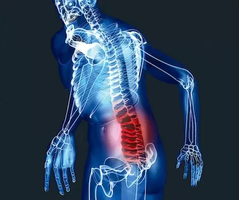
Lumbar Osteochondrosis: Symptoms and Treatment
The cause of lumbar osteochondrosis is unknown. The most important is genetic susceptibility, age-related changes in the intervertebral discs. Clumsy movements, prolonged forced postures, lifting and carrying heavy objects, exercise overload, and being overweight can all cause pain.
Depending on the duration, there are acute pain lasting up to 4 weeks, subacute (4 to 12 weeks), and chronic (lasting more than 12 weeks).
Neurological complications of lumbar osteochondrosis:
The first stage. Clinical manifestations are related to reflex muscle tone.
Low back pain (low back pain). Acute pain in the lumbar region that starts suddenly and is caused by small movements of the back. The lumbar spine has a very limited range of motion and there is compensatory scoliosis. "Stone" density of the paravertebral muscles. Low back pain lasting no more than 7-10 days with adequate treatment and lumbar immobilization.
Low back pain (back pain).Patients complain of moderate pain in the lower back, aggravated by exercise or in certain positions, and discomfort when standing or sitting for long periods of time. Onset is usually gradual. Clinically, limited lumbar mobility, paraspinal muscle tension, and soreness are often identified. In most cases, the pain will subside within 2-3 weeks, but it can become chronic pain if left untreated.
Lumboischialgia (lower back pain radiating to the legs). In the lower back, movement is limited, paraspinal muscles are tense and painful on palpation.
In piriformis syndrome, the sciatic nerve is compressed, causing paresthesia and numbness in the legs and feet. Positive Lasseger syndrome. But there were no signs of radiculopathy.
second stageNeurological complications of lumbar osteochondrosis.
A herniated disc with radiculopathy or radiculopathy. Compression at the root is accompanied by shooting in the leg, burning pain. The pain is aggravated by movement, coughing, and is accompanied by numbness of the roots, muscle weakness, and loss of reflexes. Positive stress symptoms.
In the lumbar region, the greatest load falls on the lower part, therefore, the L5 and S1 roots are most often involved in the pathological process. Each root has its own distribution of pain and numbness in the extremities.
A neurologist detected radiculopathy during an objective examination.
Lumbar Osteochondrosis Stage III Nervous System Disease.
Vascular-nerve root conflict. Paralytic sciatica syndrome occurs when blood circulation in the radicular artery L5 is disturbed, but less so in S1. Rarely, other levels of nerve root ischemia are diagnosed.
Acute back pain can occur with irradiation along the sciatic nerve during clumsy exercise or weight lifting. Then the extensor muscles of the feet and fingers become paralyzed or paralyzed while walking, and the "spanking" (stepping) of the feet. As the patient walks, the leg is raised and thrown forward while hitting the floor with the toes.
In most cases, the paralysis resolves safely within a few weeks.
Stage IV neurological complications of lumbar osteochondrosis.
Violation of the blood supply to the spinal cord and cauda equina. In spinal stenosis, several spinal nerve roots (cauda equina) are affected. Pain is mild at rest, but intermittent claudication syndrome occurs when walking. Pain that spreads from the base of the lower back to the feet when walking, accompanied by weakness, paresthesia, and numbness in the legs, which disappears at rest or when the trunk is leaned forward.
Acute spinal cord circulation disorder is the most serious complication of lumbar osteochondrosis. Acute lower extremity paralysis or paralysis. Leg weakness with lower extremity numbness, pelvic organ dysfunction.
Examination of patients with lumbar osteochondrosis.
It is very important to analyze complaints and medical records to rule out serious pathology. Neurological examination is performed to rule out damage to the roots and spinal cord. Manual examination allows you to determine the source of pain, limitation of motion, muscle spasms.
Other tests are available for suspected specific back pain.
X-rays of the lumbar spine to rule out tumors, spinal injuries, spondylolisthesis. X-ray signs of osteochondrosis have no clinical value, as all elderly and elderly people have them. A functional X-ray is done to look for spinal instability. Photos were taken in extreme flexion and extension.
For nerve root or spinal symptoms, an MRI or CT scan of the lumbar spine is required. On MRI, the herniated disc and spinal cord can be better seen, while on CT, the bone structure can be better seen. The clinical level of the lesion and the MRI findings should correspond to each other, as a herniated disc detected on MRI is not always the cause of pain.
In neurological deficits, neuroelectromyography (ENMG) is sometimes prescribed to confirm the diagnosis.
If somatic pathology is suspected, perform a thorough clinical examination.
Osteochondrosis of the lumbar spine, treatment.
At the first sign of discomfort in the lumbar spine, regular gymnastics can strengthen muscles with corsets, swimming and massage sessions.
The treatment of lumbar osteochondrosis is divided into 3 phases: acute phase, subacute phase and chronic phase.
In the acute phase, the first task is to relieve the pain syndrome as soon as possible and restore the patient's quality of life. In cases of severe pain, it is recommended to immobilize the lumbar spine with a special anti-radiculitis corset for 2-3 weeks. Bed rest should not exceed 2-3 days. In many patients, pain syndrome may be added in the context of the expansion of exercise therapy. Patients should not limit themselves to reasonable physical activity.
Among non-drug treatments, interstitial electrical stimulation, acupuncture, leech therapy, and massage are effective. Manual healing is available, but only in competent hands.
medical treatement. In acute pain, NSAIDs are required. In combination with anti-inflammatory drugs, muscle relaxants can be prescribed in the short term.
In lumbar osteochondrosis, therapeutic blockade with local anesthetics, non-steroidal anti-inflammatory drugs, and corticosteroids is effective. The drug mixture is placed as close as possible to the focal point of pain (into the affected muscle, the root's exit point).
For radiculopathy with neuropathic pain, anti-inflammatory drugs are ineffective, in which case antidepressants, anticonvulsants, and special treatment patches are prescribed.
With paralysis, numbness, vascular preparations, B vitamins are prescribed.
For prolonged myofascial pain, relaxation following the introduction of NSAIDs, muscle relaxants, acupuncture, and isometric contractions at the trigger point is effective.
For chronic pain, antidepressants, exercise therapy, and other nonpharmacological treatments are the treatment of choice.
Spinal stenosis, weight loss, wearing a corset, NSAIDs, and various IVs are all required.
Surgical treatment of paralyzed sciatica (first three days) and cauda equina syndrome (tetraplegia, impaired sensitivity, urinary and fecal incontinence).
Prevention of lumbar osteochondrosis
preventionlumbar osteochondrosisReduce to avoid prolonged, uncomfortable positions, overloading. It is important to properly equip your workplace, alternating work and rest periods. Wear a harness to prevent overloading your body. Do some exercises to strengthen your back muscles.